

Non-Invasive Cerebral Hemodynamics, Vasospasm Surveillance & Neonatal Neurosonography
Neuro-Ultrasound, Transcranial Doppler (TCD) & Carotid Duplex
Clinical Overview & Pathophysiology
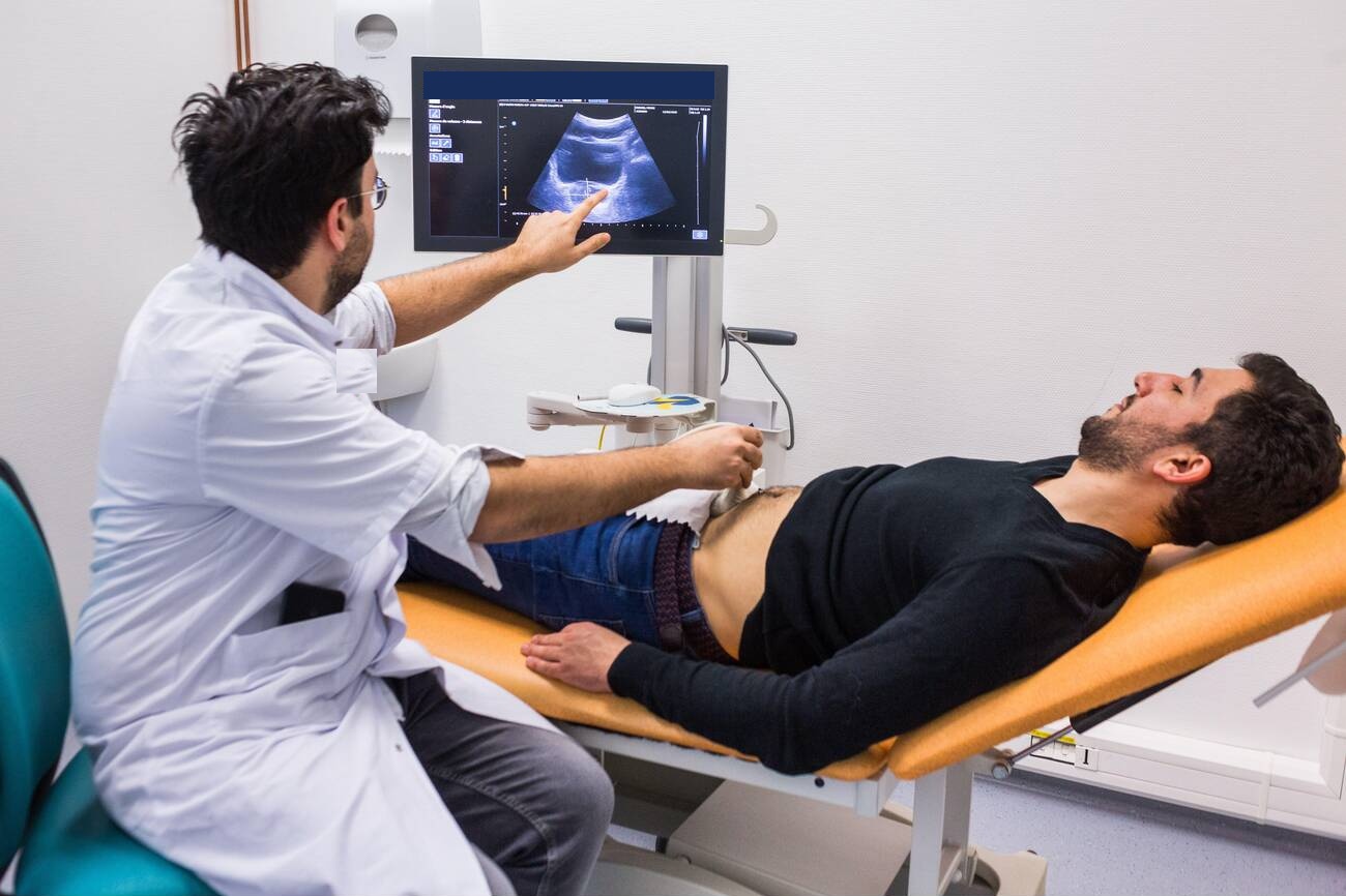
Ultrasonography and Doppler imaging provide rapid, radiation-free, real-time hemodynamic and anatomical evaluation of intracranial and extracranial cerebral circulation and neural structures. Transcranial Doppler (TCD) is the premier non-invasive modality for measuring blood flow velocity in the basal cerebral arteries through acoustic bone windows (transtemporal, transorbital, suboccipital), serving as an indispensable daily monitor for cerebral vasospasm following aneurysmal subarachnoid hemorrhage. Extracranial Carotid and Vertebral Duplex Doppler quantifies atherosclerotic plaque burden and stenosis to prevent ischemic strokes. In addition, Intraoperative Ultrasound (iUS) provides real-time parenchymal imaging during craniotomies, while Neonatal Cranial Ultrasound through open fontanelles detects intraventricular hemorrhage and hydrocephalus without requiring sedation. Dr. Ashok Kumar utilizes advanced neuro-ultrasound across diagnostic, intensive care, and surgical settings.
Clinical Classifications & Pathological Subtypes
- Transcranial Doppler (TCD) & TCD Monitoring: Non-invasive pulsed 2 MHz Doppler measuring mean flow velocity (MFV) and pulsatility index (PI) in the Middle, Anterior, and Posterior Cerebral Arteries and Basilar Artery.
- Carotid & Vertebral Duplex Doppler Ultrasound: High-resolution B-mode imaging combined with color and spectral Doppler quantifying internal carotid artery stenosis, plaque ulceration, and vertebral flow reversal.
- Intraoperative Neurosurgical Ultrasound (iUS): Specialized micro-convex surgical transducers placed directly onto the exposed dura or brain cortex during tumor resection to compensate for brain shift and verify complete removal.
- Neonatal Cranial Neurosonography: Bedside imaging through the anterior fontanelle assessing ventricular dilation (hydrocephalus), periventricular leukomalacia (PVL), and germinal matrix hemorrhage.
- Optic Nerve Sheath Diameter (ONSD) Ultrasound: Bedside ocular ultrasound measuring the retrobulbar optic nerve sheath diameter as a non-invasive surrogate metric of raised intracranial pressure (ICP).
Key Symptoms & Clinical Presentation
!Emergency Red Flags & Immediate Surgical Indications
TCD middle cerebral artery mean flow velocity exceeding 200 cm/s with a Lindegaard Ratio > 6 (indicating severe critical cerebral vasospasm requiring emergent hemodynamic augmentation or intra-arterial spasmolysis) or unstable, ulcerated, hypoechoic carotid plaques with > 80% stenosis. Immediate neurosurgical intervention is vital.
Diagnostic & Neuro-Evaluation Workflow
Multi-Frequency Digital Color Duplex Ultrasound Systems
Equipped with dedicated linear vascular probes (5-12 MHz) and phased-array low-frequency sector transducers (1-3 MHz).
Transcranial Power Doppler with M-Mode
Simultaneous depth-gate recording tracing blood flow spectra across multiple intracranial vessel depths simultaneously.
Contrast-Enhanced Ultrasound (CEUS)
Microbubble contrast agent infusion for high-resolution microvascular perfusion imaging of brain tumors and plaque neovascularization.
Non-Invasive Intracranial Compliance Profiling
Continuous waveform analysis calculating resistivity index (RI) and pulsatility index (PI) to assess cerebral microvascular resistance.
Advanced Treatments & Procedures by Dr. Ashok Kumar
Vasospasm-Guided Hyperdynamic Therapy
Guiding induced hypertension, euvolemia, and intra-arterial nimodipine infusions in the Neuro-ICU based on daily TCD velocity metrics.
Carotid Endarterectomy (CEA) Selection
Identifying symptomatic and asymptomatic patients meeting NASCET criteria for microsurgical carotid plaque removal to prevent catastrophic strokes.
Real-Time Resection Guidance in Brain Surgery
Using iUS to visualize deep-seated tumor margins, identify the ventricle, and confirm empty tumor resection cavities before closing the dura.
Fontanelle Tap & Ventricular Access Guidance
Real-time ultrasound guidance for bedside reservoir needle placement or ventricular puncture in neonates.
Post-Operative Recovery & Long-Term Prognosis
Neuro-ultrasound procedures are completely safe, non-invasive, radiation-free, and pain-free, requiring zero needles or sedation. Patients can undergo repeated scans at the bedside in the ICU or outpatient clinic without any recovery time.
Frequently Asked Questions (Clinical FAQs)
Authoritative, medically verified answers to critical clinical questions regarding symptoms, surgical safety, recovery timelines, and long-term prognosis.
What is Transcranial Doppler (TCD) and how does it prevent brain damage after an aneurysm bleed?
How does Carotid Duplex Ultrasound detect arterial blockages that cause strokes?
What is Intraoperative Ultrasound (iUS) and how does it assist Dr. Ashok Kumar during brain surgery?
Why is cranial ultrasound the safest initial test for newborns with suspected hydrocephalus?
Is there any radiation, contrast dye, or discomfort involved in neuro-ultrasound examinations?
Neuro-Diagnostics & Advanced Imaging
- Advanced High-Field Neuro-MRI: Brain & Spine Protocols
- High-Speed Multi-Slice Computed Tomography (Brain & Spine CT)
- Cerebral Angiography, Digital Subtraction Angiography (DSA) & Vascular Neuro-Imaging
- Digital Spine & Cranial Radiography (Dynamic Neuro-Imaging)
- Neuro-Ultrasound, Transcranial Doppler (TCD) & Carotid Duplex
- Digital Electroencephalography (EEG) & Advanced Brain Mapping
- Comprehensive Neuro-Rehabilitation & Specialized Physiotherapy
Why Choose Dr. Ashok Kumar
Emergency 24 Hours Dr. Ashok Kumar
Neuro-Spine OPD Location
Direct Consultation & Emergency
Email: dr.ashokkumar4108@gmail.com
Consultation Hours
Emergency Neuro-Trauma: 24/7 Open
