

Ultra-Fast Trauma Neuro-Imaging, 3D Calvarial/Vertebral Reconstructions & CT Cisternography
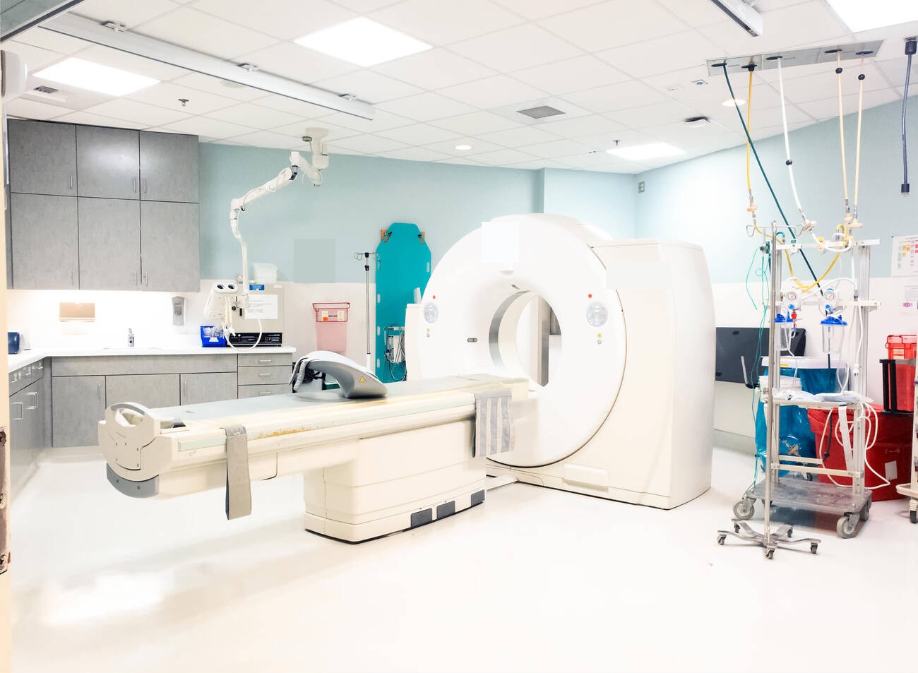
High-Speed Multi-Slice Computed Tomography (Brain & Spine CT)
Clinical Overview & Pathophysiology
Computed Tomography (CT) is the indispensable frontline imaging modality in emergency neurology and neurosurgery, delivering ultra-rapid, cross-sectional volumetric visualization of the head, skull base, and entire spinal column within seconds. Utilizing modern Multi-Detector Helical CT (MDCT) with sub-millimeter slice collimation, CT scanning provides unparalleled clarity for detecting acute intracranial hemorrhage, skull fractures, acute ischemic stroke demarcation, vertebral fractures, and osseous spinal canal compromise. Dr. Ashok Kumar utilizes advanced CT diagnostic protocols—including emergency Non-Contrast Cranial CT (NCCT), CT Angiography (CTA), CT Cisternography for CSF leaks, and 3D stereolithic bone reconstructions for complex spine trauma and craniofacial surgery.
Clinical Classifications & Pathological Subtypes
- Non-Contrast Cranial CT (NCCT Head): The emergency gold standard for rapid hyperacute detection of epidural, subdural, subarachnoid, and intraparenchymal brain hemorrhages.
- CT Angiography (CTA) of Brain & Neck: High-speed contrast-enhanced volumetric arterial imaging mapping intracranial aneurysms, arterial dissections, and large vessel occlusions (LVO) in stroke.
- Multi-Slice Spine CT with 3D Reconstructions: Sub-millimeter axial scanning with multiplanar sagittal, coronal, and 3D volume-rendered reconstructions for complex vertebral burst fractures and pedicle screw planning.
- CT Cisternography: Intrathecal non-ionic contrast injection followed by high-resolution skull base CT to pinpoint the precise anatomical breach in traumatic or spontaneous CSF rhinorrhea.
- Post-Operative Fusion & Hardware CT: High-contrast bone window imaging evaluating spinal pedicle screw position, interbody cage incorporation, and solid bony arthrodesis.
Key Symptoms & Clinical Presentation
!Emergency Red Flags & Immediate Surgical Indications
Acute respiratory depression, severe traumatic coma (GCS < 8), uncal herniation with blown pupil, and post-traumatic spinal shock. CT scanning must be executed seamlessly with trauma resuscitation teams maintaining continuous in-line spinal stabilization and airway protection.
Diagnostic & Neuro-Evaluation Workflow
High-Speed 128/256-Slice Multi-Detector Scanner
Acquires sub-millimeter (0.5 mm) volumetric isotropic data of the entire brain in less than 3 to 5 seconds, virtually eliminating patient motion artifacts.
Iterative AI-Assisted Dose Reduction (ASiR / ALARA)
Advanced reconstruction algorithms reducing patient radiation dose by up to 60-70% while enhancing diagnostic bone and soft-tissue sharpness.
3D Multi-Planar Reformation (MPR)
Real-time multi-angle reformatting enabling the surgeon to inspect complex spinal fractures from any anatomical vector.
Intravenous Non-Ionic Low-Osmolar Iodinated Contrast
Pre-warmed non-ionic contrast administered via automated power injectors for dynamic arterial timing during CT Angiography.
Advanced Treatments & Procedures by Dr. Ashok Kumar
Emergency Neuro-Trauma Protocol
Immediate bedside wet-read reporting by senior radiologist and Dr. Ashok Kumar within 10 minutes of patient arrival.
Stereotactic Neuro-Navigation Data Export
Seamless digital transfer of DICOM datasets directly to operating room surgical navigation consoles.
CT-Guided Spinal Injections
Precise needle placement for transforaminal epidural steroid injections (TFESI) and facet joint denervation blocks under direct CT guidance.
Skull Base Defect Mapping
Coronal fine-cut bone algorithms identifying micro-fractures of the cribriform plate, fovea ethmoidalis, and tegmen tympani.
Post-Operative Recovery & Long-Term Prognosis
A non-contrast head CT scan takes less than 60 seconds of actual scan time and requires no recovery period. If intravenous contrast was administered, patients are observed for 15 to 30 minutes, encouraged to drink fluids to promote contrast clearance, and discharged immediately.
Frequently Asked Questions (Clinical FAQs)
Authoritative, medically verified answers to critical clinical questions regarding symptoms, surgical safety, recovery timelines, and long-term prognosis.
Why is a CT scan preferred over an MRI as the first test in acute head injury?
How much radiation is involved in a brain CT, and is it dangerous?
When is an MRI necessary if a patient has already had a normal CT scan?
What is CT Cisternography and how does it locate a brain fluid (CSF) leak?
What precautions should be taken before undergoing a Contrast-Enhanced CT scan?
Neuro-Diagnostics & Advanced Imaging
- Advanced High-Field Neuro-MRI: Brain & Spine Protocols
- High-Speed Multi-Slice Computed Tomography (Brain & Spine CT)
- Cerebral Angiography, Digital Subtraction Angiography (DSA) & Vascular Neuro-Imaging
- Digital Spine & Cranial Radiography (Dynamic Neuro-Imaging)
- Neuro-Ultrasound, Transcranial Doppler (TCD) & Carotid Duplex
- Digital Electroencephalography (EEG) & Advanced Brain Mapping
- Comprehensive Neuro-Rehabilitation & Specialized Physiotherapy
Why Choose Dr. Ashok Kumar
Emergency 24 Hours Dr. Ashok Kumar
Neuro-Spine OPD Location
Direct Consultation & Emergency
Email: dr.ashokkumar4108@gmail.com
Consultation Hours
Emergency Neuro-Trauma: 24/7 Open
