

Distinguishing Central vs Peripheral Vertigo, Resection of Acoustic Neuromas & Cerebellar Lesions
Dizziness, Vertigo & Posterior Fossa Neuro-Evaluation
Clinical Overview & Pathophysiology
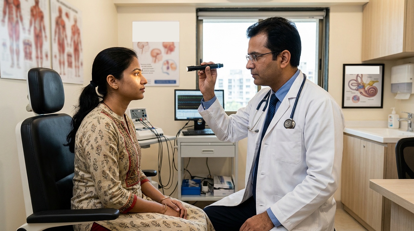
Dizziness, true rotatory vertigo, and disequilibrium are among the most frequent yet diagnostically challenging clinical complaints. While benign peripheral vestibulopathies (such as BPPV and vestibular neuritis) originate in the inner ear, central vertigo represents serious intracranial pathology situated within the posterior cranial fossa, cerebellum, or brainstem. Pathologies include Acoustic Neuromas (Vestibular Schwannomas), Meningiomas of the Cerebellopontine Angle (CPA), Posterior Fossa Infarctions, Vertebrobasilar Insufficiency, and Chiari Malformations. Dr. Ashok Kumar provides meticulous neuro-otological differentiation, advanced vestibular diagnostics, and high-precision microsurgery.
Clinical Classifications & Pathological Subtypes
- Central Vertigo: Vertigo arising from pathology in the vestibular nuclei, brainstem, or cerebellum, characterized by constant symptoms and neurological signs.
- Cerebellopontine Angle (CPA) Tumors: Acoustic neuromas (Vestibular Schwannomas) and CPA meningiomas compressing cranial nerves VII, VIII, and the brainstem.
- Posterior Fossa Stroke & TIA: Ischemic or hemorrhagic injury to the cerebellar hemispheres or PICA/AICA territories producing acute vertigo.
- Chiari Malformation Type I: Cerebellar tonsillar herniation through the foramen magnum impinging on the brainstem and vestibular pathways.
- Vestibular Paroxysmia: Microvascular compression of the 8th cranial nerve root producing brief, frequent recurring bursts of vertigo.
Key Symptoms & Clinical Presentation
!Emergency Red Flags & Immediate Surgical Indications
The HINTS examination showing central patterns (Normal head impulse test, direction-changing nystagmus, Test of skew deviation), acute inability to sit or stand unsupported, sudden onset facial numbness, dysarthria, or dysphagia. This pattern indicates acute cerebellar stroke or hemorrhage.
Diagnostic & Neuro-Evaluation Workflow
High-Resolution Contrast 3T MRI of Internal Auditory Canal (IAC)
Sub-millimeter thin T1-contrast and 3D CISS/FIESTA sequences capable of visualizing intracanalicular tumors as small as 1 to 2 mm.
MR Angiography of Vertebrobasilar Circulation
Detailed 3D reconstruction evaluating vertebral artery stenosis, PICA/AICA anatomy, and basilar dolichoectasia.
Brainstem Auditory Evoked Potentials (BAEP / BERA)
Electrophysiological testing measuring inter-peak latencies (I-III and I-V) to detect retrocochlear conduction delays along CN VIII.
Pure Tone Audiometry (PTA) & Speech Discrimination
Quantitative acoustic thresholds measuring sensorineural deficit and speech clarity reduction.
Advanced Treatments & Procedures by Dr. Ashok Kumar
Retrosigmoid Keyhole Microsurgery for Acoustic Neuroma
Skull base approach utilizing high-power operative microscopy and continuous facial nerve (CN VII) monitoring to preserve facial movement and hearing.
Translabyrinthine & Middle Cranial Fossa Approaches
Tailored anatomical corridors chosen according to tumor size, extension, and pre-existing auditory function.
Posterior Fossa Craniectomy & Decompression
Suboccipital decompression with duroplasty for Chiari malformations and large cerebellar compressive masses.
Microvascular Decompression of CN VIII
Micro-neurosurgical separation of compressing arterial loops causing refractory vestibular paroxysmia.
Post-Operative Recovery & Long-Term Prognosis
Following acoustic neuroma or posterior fossa surgery, patients mobilize within 48 to 72 hours under specialized vestibular physiotherapist supervision. Central vestibular compensation adapts over 4 to 12 weeks, restoring confidence in balance, walking, and head movements.
Frequently Asked Questions (Clinical FAQs)
Authoritative, medically verified answers to critical clinical questions regarding symptoms, surgical safety, recovery timelines, and long-term prognosis.
How can a patient tell the difference between common ear vertigo and a brain tumor?
Can facial expression and smile be preserved during Acoustic Neuroma surgery?
What is the HINTS exam, and why is it superior to a brain CT in acute vertigo?
What are the treatment options if an Acoustic Neuroma is detected while still very small?
What is Vestibular Rehabilitation Therapy (VRT) and how does it help?
Brain Surgery & Cranial Conditions
- Brain Tumor Surgery & Neuro-Oncology
- Hydrocephalus Management: VP Shunt & ETV Surgery
- Head Injury & Emergency Neuro-Trauma Care
- Meningitis & Central Nervous System Infections
- Headache & Cranial Neuralgias: Neurosurgical Care
- Dizziness, Vertigo & Posterior Fossa Neuro-Evaluation
- Epilepsy & Seizure Disorders: Neurosurgical Evaluation
Why Choose Dr. Ashok Kumar
Emergency 24 Hours Dr. Ashok Kumar
Neuro-Spine OPD Location
Direct Consultation & Emergency
Email: dr.ashokkumar4108@gmail.com
Consultation Hours
Emergency Neuro-Trauma: 24/7 Open
