

3T MRI, Diffusion Tensor Imaging (DTI Tractography), MR Spectroscopy & Functional MRI
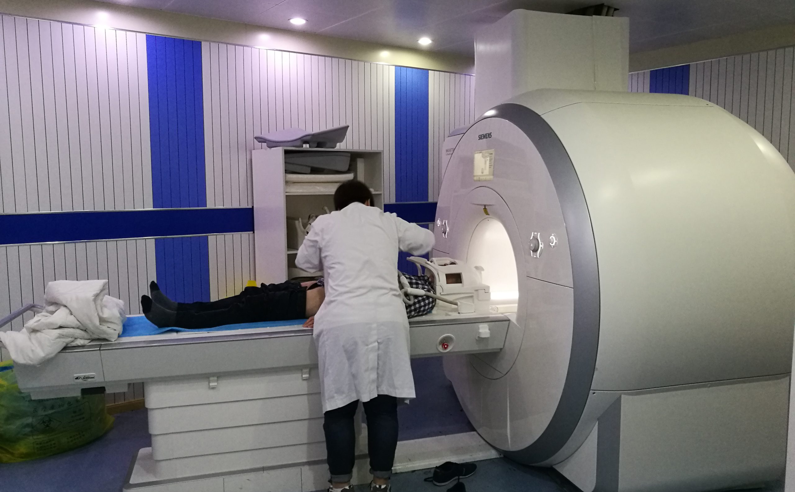
Advanced High-Field Neuro-MRI: Brain & Spine Protocols
Clinical Overview & Pathophysiology
Magnetic Resonance Imaging (MRI) is the premier, non-invasive imaging modality for visualizing central and peripheral nervous system pathology, offering unmatched soft-tissue contrast resolution without exposing patients to ionizing radiation. Utilizing high-field 3.0 Tesla magnet technology and multi-channel phased-array head and spine coils, Dr. Ashok Kumar’s diagnostic protocols provide sub-millimeter visualization of the cerebral cortex, subcortical nuclei, brainstem, cranial nerves, spinal cord parenchyma, intervertebral discs, and thecal sac. Advanced functional sequences—including MR Spectroscopy (MRS), Diffusion Tensor Imaging (DTI), Functional MRI (fMRI), and Phase-Contrast CSF Flow Cine—allow definitive biochemical characterization of lesions and preoperative mapping of eloquent motor and language pathways.
Clinical Classifications & Pathological Subtypes
- Multi-Parametric 3T Brain MRI: Axial, sagittal, and coronal T1-weighted (with and without Gadolinium contrast), T2-weighted, FLAIR, and Susceptibility-Weighted Imaging (SWI).
- Advanced MR Spectroscopy (MRS): In-vivo non-invasive biochemical analysis measuring Choline, Creatine, NAA, Myo-inositol, and Lipid-Lactate peaks to differentiate tumors from radiation necrosis or abscesses.
- Diffusion Tensor Imaging (DTI Tractography): 3D vector mapping of white matter tracts (corticospinal tract, arcuate fasciculus) integrated directly into intraoperative neuro-navigation.
- Functional MRI (BOLD fMRI): Blood-Oxygen-Level-Dependent activation mapping identifying primary motor cortex, sensory strips, and Broca/Wernicke language dominance.
- High-Definition Whole-Spine MRI: Multi-segment profiling of cervical, thoracic, and lumbosacral regions evaluating disc herniations, cord myelopathy, syrinx cavities, and spinal cord tumors.
Key Symptoms & Clinical Presentation
!Emergency Red Flags & Immediate Surgical Indications
Absolute contraindications to MRI scanning: Non-MRI-conditional cardiac pacemakers, implantable cardioverter-defibrillators (ICDs), metallic foreign bodies in the orbits, ferromagnetic cerebral aneurysm clips, and non-conditional cochlear implants. Mandatory pre-scan safety screening is conducted for every patient.
Diagnostic & Neuro-Evaluation Workflow
High-Field 3-Tesla (3T) Superconducting Magnet
Delivers double the signal-to-noise ratio of conventional 1.5T scanners, enabling ultra-fast scan times and sub-millimeter anatomical detail.
Multi-Channel Digital Phased-Array Neuro Coils
Dedicated 32-channel head and whole-spine coils optimizing signal reception across the brain and spinal canal.
Gadolinium-Based Contrast Enhancement (GBCA)
Macrocyclic, highly stable contrast agents utilized to delineate breakdown of the blood-brain barrier in tumors, infections, and active MS plaques.
Renal Function & eGFR Safety Protocol
Serum creatinine and estimated Glomerular Filtration Rate (eGFR) verification prior to contrast injection to prevent Nephrogenic Systemic Fibrosis (NSF).
Advanced Treatments & Procedures by Dr. Ashok Kumar
Epilepsy Protocol MRI
Specialized thin-slice oblique coronal sequences angled perpendicular to the hippocampus for detecting subtle hippocampal sclerosis and focal cortical dysplasias.
Pituitary Dynamic Micro-Adenoma MRI
Rapid dynamic post-contrast imaging capturing the differential contrast wash-in between normal adenohypophysis and hypoperfused microadenomas.
Internal Auditory Canal (IAC) 3D CISS/FIESTA
Sub-millimeter cisternographic sequences isolating the 7th and 8th cranial nerves in the cerebellopontine angle.
Phase-Contrast CSF Flow Cine Studies
Physiological velocity-encoded mapping quantifying CSF stroke volume across the Aqueduct of Sylvius and Foramen Magnum in Chiari and NPH.
Post-Operative Recovery & Long-Term Prognosis
MRI is a completely painless outpatient procedure lasting 20 to 45 minutes depending on protocol complexity. Patients can resume routine activities immediately. A formal detailed neuro-radiological report correlated with Dr. Ashok Kumar's clinical neurosurgical findings is provided for comprehensive consultation.
Frequently Asked Questions (Clinical FAQs)
Authoritative, medically verified answers to critical clinical questions regarding symptoms, surgical safety, recovery timelines, and long-term prognosis.
Why is a 3-Tesla (3T) MRI superior to older 1.5T scanners for brain and spine conditions?
What is MR Spectroscopy (MRS) and how does it differentiate a brain tumor from an infection?
How does Diffusion Tensor Imaging (DTI Tractography) prevent paralysis during surgery?
Is MRI contrast (Gadolinium) safe, and who requires a kidney function test?
How are patients with claustrophobia or anxiety accommodated during an MRI scan?
Neuro-Diagnostics & Advanced Imaging
- Advanced High-Field Neuro-MRI: Brain & Spine Protocols
- High-Speed Multi-Slice Computed Tomography (Brain & Spine CT)
- Cerebral Angiography, Digital Subtraction Angiography (DSA) & Vascular Neuro-Imaging
- Digital Spine & Cranial Radiography (Dynamic Neuro-Imaging)
- Neuro-Ultrasound, Transcranial Doppler (TCD) & Carotid Duplex
- Digital Electroencephalography (EEG) & Advanced Brain Mapping
- Comprehensive Neuro-Rehabilitation & Specialized Physiotherapy
Why Choose Dr. Ashok Kumar
Emergency 24 Hours Dr. Ashok Kumar
Neuro-Spine OPD Location
Direct Consultation & Emergency
Email: dr.ashokkumar4108@gmail.com
Consultation Hours
Emergency Neuro-Trauma: 24/7 Open
