

Intradural Extramedullary, Intramedullary & Vertebral Tumor Resection with Instrumented Spinal Stabilization
Spine Tumor Surgery & Spinal Oncology
Clinical Overview & Pathophysiology
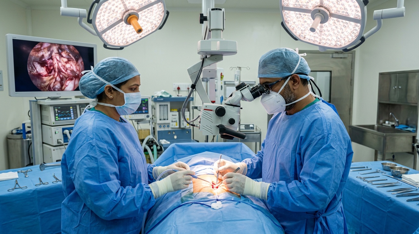
Spinal tumors are abnormal neoplasms arising within or adjacent to the spinal cord, nerve roots, meningeal coverings, or vertebral osseous column. They are classified into three anatomical compartments: Intradural Extramedullary tumors (Schwannomas, Neurofibromas, Meningiomas), Intramedullary Spinal Cord Tumors (Ependymomas, Astrocytomas, Hemangioblastomas occurring directly inside the neural tissue), and Extradural / Vertebral Metastases (arising from breast, lung, prostate, or renal primaries). Spinal tumors pose a severe risk of irreversible quadriplegia or paraplegia due to direct mechanical compression of ascending and descending spinal tracts. Dr. Ashok Kumar utilizes high-power micro-neurosurgery, continuous intraoperative neuro-monitoring (MEP/SSEP/D-Wave), and 3D spinal reconstruction to achieve safe tumor excision and preserve spinal alignment.
Clinical Classifications & Pathological Subtypes
- Intradural Extramedullary Tumors: Benign sheath tumors (Schwannomas, Neurofibromas) and Meningiomas located inside the dura but outside the spinal cord.
- Intramedullary Spinal Cord Tumors: Primary tumors developing within the spinal cord substance (Ependymomas, Astrocytomas, Hemangioblastomas) requiring midline myelotomy.
- Extradural / Metastatic Spine Tumors: Malignant secondary deposits infiltrating the vertebral body and pedicles, frequently precipitating pathological compression fractures.
- Primary Vertebral Bone Neoplasms: Giant cell tumors, chordomas, osteosarcomas, and plasmacytomas requiring en-bloc or radical corpectomy.
- Spinal Dumbbell Neoplasms: Tumors extending both inside the spinal canal and outside into the thoracic or retroperitoneal cavity through an enlarged neural foramen.
Key Symptoms & Clinical Presentation
!Emergency Red Flags & Immediate Surgical Indications
Rapidly ascending motor weakness within 24-48 hours, sudden urinary retention with overflow incontinence, saddle anesthesia, and bilateral foot drop. This constitutes an oncological emergency requiring immediate high-dose corticosteroids, urgent contrast MRI, and emergent surgical decompression.
Diagnostic & Neuro-Evaluation Workflow
Whole-Spine Contrast 3T MRI with STIR
High-resolution sagittal and axial T1 with gadolinium, T2, and Short Tau Inversion Recovery (STIR) sequences assessing cord edema and intradural tumor boundaries.
Thin-Slice CT Spine with 3D Bone Reconstructions
Detailed assessment of pedicle osteolysis, vertebral body collapse, and Spinal Instability Neoplastic Score (SINS).
CT-Guided Percutaneous Needle Biopsy
Minimally invasive diagnostic histological confirmation of suspected metastatic lesions prior to definitive surgical planning.
Whole-Body 18F-FDG PET-CT
Comprehensive staging to identify primary occult malignancy and detect additional systemic metastatic foci.
Advanced Treatments & Procedures by Dr. Ashok Kumar
Microsurgical Intradural Tumor Resection
Laminotomy or laminectomy with high-magnification microsurgical dissection along arachnoid planes to resect meningiomas and schwannomas completely.
Midline Posterior Myelotomy for Intramedullary Tumors
Careful micro-dissection through the posterior median sulcus of the spinal cord with continuous D-wave monitoring to excise ependymomas.
Corpectomy & Vertebral Body Replacement
Surgical removal of diseased vertebral body and placement of an expandable titanium mesh cage packed with autologous bone graft.
Instrumented Multi-Level Pedicle Screw Fixation
Rigid spinal reconstruction using titanium screws and rods to restore mechanical stability and allow immediate post-operative ambulation.
Percutaneous Vertebroplasty / Kyphoplasty
Minimally invasive cement augmentation for painful metastatic compression fractures without spinal cord impingement.
Post-Operative Recovery & Long-Term Prognosis
Patients are mobilized on post-operative day 2 with a custom rigid spinal orthosis (brace). Continuous neuro-rehabilitation begins immediately in the hospital. Multidisciplinary referral is coordinated with Radiation Oncology for targeted Stereotactic Body Radiotherapy (SBRT) for metastatic lesions.
Frequently Asked Questions (Clinical FAQs)
Authoritative, medically verified answers to critical clinical questions regarding symptoms, surgical safety, recovery timelines, and long-term prognosis.
Can a benign spinal tumor cause permanent paralysis if not removed?
How does Dr. Ashok Kumar prevent spinal cord damage during tumor removal?
What is the Spinal Instability Neoplastic Score (SINS)?
What is the difference between a Laminectomy and a Laminoplasty for tumor exposure?
How soon can a patient walk after spinal tumor surgery?
Spine Surgery & Spinal Disorders
Why Choose Dr. Ashok Kumar
Emergency 24 Hours Dr. Ashok Kumar
Neuro-Spine OPD Location
Direct Consultation & Emergency
Email: dr.ashokkumar4108@gmail.com
Consultation Hours
Emergency Neuro-Trauma: 24/7 Open
